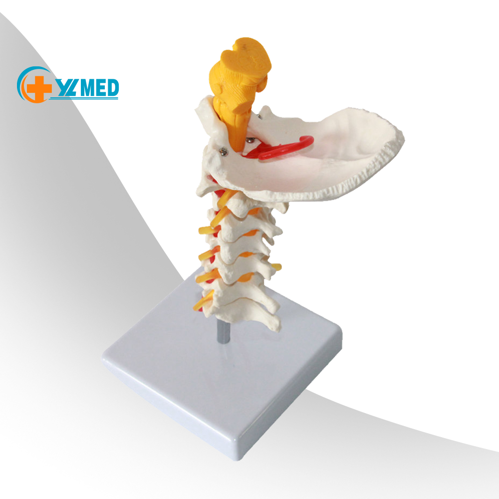
Bone at the back of the skull that protects the brain
The occipital bone is a flat, trapezoid-shaped bone that houses the back part of the brain. It is located at the lower back of the cranium and is one of seven bones that form your skull. Biology Prepared Slides and Accessories
This article will review the structure and function of the occipital bone of the skull, as well as problems that can affect the bone.
The occipital bone has many important functions, but its most important role is protecting your brain. It also supports the muscles of your neck. The occipital bone protects the brain’s visual processing center and acts as the connecting pathway from the brain to the spine.
As the occipital bone connects with the first vertebra (atlas), it forms the atlantooccipital joint. This junction helps you to nod and shake your head. The atlas is also the direct link between the spine and the skull.
Given its location, the occipital bone affects all your body’s movements, as well as your flexibility, stability, and balance. It also plays a part in your ability to see and interact with the world around you.
The occipital bone is located at the lower back of the head and connects to the spine and five of the other bones in the cranium.
As you get older, your occipital bones will fuse to the other bones of your skull. Your sphenoid bone, which is located in the middle of your skull, will fuse with the occipital bone when you are between the ages of 18 and 25. Then, between the ages of 26 and 40, the parietal bones at the top of your head and the occipital bone will fuse together.
At the base of your skull, there is a large oval opening in the occipital bone that is called the foramen magnum. This opening allows for the passage of the spinal cord. The occipital bone is the only cranial bone to connect to the cervical spine.
Like other bones in your skull, the occipital bone is flat, and it has many attachments and features, which is why it is often described in parts.
The foramen magnum is curved externally and hollow inside. It is the passageway of the central nervous system through the skull that connects the brain to the spinal cord. The structures that pass through the foramen magnum include:
The foramen magnum is divided into four parts: one basilar part, two condylar parts, and a squamous part. All four are part of the opening of the foramen magnum.
The basilar part of the occipital bone is at the front of the foramen magnum and sits next to the dense area of the temporal bone of your skull surrounding the inner ear.
Toward the front, the basilar part fuses to the sphenoid bone and forms the tribasilar bone. This fusion happens during puberty. The pharyngeal tubercle leading to the airway (pharynx) is on the lower surface of the basilar part.
The two condylar parts are located adjacent to the foramen magnum. They are oval-shaped and connect to the first cervical vertebra.
Next to these parts are the condylar canals where the condylar emissary veins connect the external vertebral venous plexuses to the sigmoid sinuses. The hypoglossa l nerve (the 12th cranial nerve) goes through the condylar part of the occipital bone.
The squamous part is the largest part of the occipital bone. It is located above and behind the foramen magnum and curved downward on each side.
There are two curved lines on each side of the squamous part: the highest nuchal line and the superior nuchal line. There is also a middle line running through the nuchal plane (inferior nuchal line ). The nuchal plane is rough and irregular and attaches to several muscles of the head and neck.
The internal surface of the squamous part is bowl-shaped and divided into four depressions of the cruciform eminence:
Along the internal surface of the occipital bone, there is a point of intersection of the four divisions of the cruciform eminence called the internal occipital protuberance. It runs from the superior angle of the bone to a deep groove (sagittal sulcus ), which hides part of the superior sagittal sinus and is attached to the falx cerebri .
The superior sagittal sinus allows blood to drain from the adjacent parts of the anterior hemisphere to the sinuses. At the upper part is the internal occipital crest is the transverse sinuses. The union of the transverse and sagittal sinuses is noted by a depression on either side of the protuberance.
The external occipital protuberance is a slight bump located at the back of your skull, just above your neck. Some people, especially males, may report an enlarged one that can be felt. This is called an occipital spur or occipital knob (or sometimes, a "knowledge bump"). A large occipital protuberance is considered normal, though people who experience pain related to an occipital spur may choose to have it reduced surgically.
Any problems with the development of occipital bone can lead to health problems. For example, if the occipital bone is misaligned, the spine may also be out of alignment which can lead to pain.
The occipital bone is sensitive and can get injured or damaged during childbirth. The bone can also be affected by traumas or injuries, such as automobile accidents, sports injuries, and falls, resulting in chronic health conditions and mental health issues (like depression and anxiety) if a traumatic brain injury occurs.
When the occipital bone is functioning or moving incorrectly, it can lead to problems with sensory processing, pain in the back of the head, neck, shoulders, or back, and lowered immune function.
Some other examples of problems that can be related to the occipital bone include vision problems, headaches, and dizziness/balance problems.
Damage to the occipital bone, such as from a fracture, can lead to vision problems or even blindness. Defects in the occipital bone can also cause severe problems with vision.
Knobloch syndrome is a type of genetic condition that is caused by a defect in the occipital bone. In this disorder, a defect in the occipital bone causes severe nearsightedness or blindness.
Occipital migraines are moderate to severe headaches that are felt in the back of the head. The cause of this type of headache is not well understood. Problems with the occipital bone can cause other types of headaches, too.
A malformation of the occipital bone is also associated with Chiari malformation, a rare condition where part of the brain protrudes through the opening in the occipital bone. This condition can cause many symptoms, including an occipital headache.
Rarely, non-cancerous (benign) bone cysts can form in the occipital bone, which can cause headaches. These cysts can usually be removed surgically.
A condition called occipital neuralgia may happen after an accident where a person had an impact on the back of their head. This condition can cause occipital pain similar to an occipital migraine, and can also lead to other symptoms like vertigo and sensitivity to light.
Occipital neuralgia can lead to neck pain and difficulty with balance and coordination.
A fractured occipital bone can be from accidents and injuries. Antibiotics or surgery might be needed if there are skin breaks or deep indentations in the bone. Fractures at the base of the occipital bone need to be closely watched for signs of nerve damage and/or meningitis.
If a person also has a traumatic brain injury, permanent neurological damage or even death can happen if it is not treated.
As your brain’s protector, your occipital bone plays an important role in your overall health and quality of life. It’s important to find out the cause of head and spine symptoms you may have, especially if you have pain and problems with your body’s function and movement.
Your healthcare provider can recommend treatments, from medications to physical therapy and surgery, depending on the underlying cause that is determined.
Physical therapy for occipital bone problems can correct misalignment and help manage pain and may help restore function and correct body movement.
The occipital bone is at the back of your head and is one of the seven skull bones. It helps protect your brain and also helps you move your head. If you have problems with your occipital bone and have symptoms like headaches, you may need physical therapy.
Betts GJ. Anatomy and Physiology 2E. Openstax; 2022.
İlhan Bahşi, Saliha Seda Adanir, Mustafa Orhan, et al. Anatomical evaluation of the foramen magnum on cone-beam computed tomography images and review of literature. Published online November 8, 2021. doi:10.7759/cureus.19385
Varghese E Dr, Samson RS Dr, Kumbargere SN Dr, Pothen M Dr. Occipital spur: understanding a normal yet symptomatic variant from orthodontic diagnostic lateral cephalogram. BMJ Case Rep. 2017:bcr2017220506. doi:10.1136/bcr-2017-220506
MSKTC. Changes in emotion after traumatic brain injury.
London Health Sciences Centre. Basal skull fracture.
National Library of Medicine. Knobloch syndrome.
Baldelli I, Mangialardi ML, Salgarello M, Raposio E. Peripheral occipital nerve decompression surgery in migraine headache. Plast Reconstr Surg Glob Open. 2020;8(10). doi:10.1097/GOX.0000000000003019
National Organization for Rare Disorders. Chiari malformations.
American Association of Neurological Surgeons. Occipital neuralgia.
UMass Chan. What is a TBI?.
American Association of Neurological Surgeons. Traumatic brain injury.
By Lana Barhum Lana Barhum has been a freelance medical writer since 2009. She shares advice on living well with chronic disease.
Thank you, {{form.email}}, for signing up.
There was an error. Please try again.
Skull Diagram By clicking “Accept All Cookies”, you agree to the storing of cookies on your device to enhance site navigation, analyze site usage, and assist in our marketing efforts.